Investigation of bone material properties

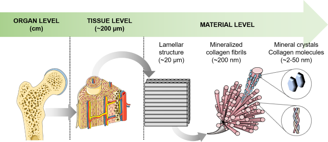
The bone matrix is composed mainly of three components represented by a mineral phase composed of poorly crystalline carbonated hydroxyapatite, an organic phase composed mostly of type I collagen and water. The HiMolA platform has developed over years a strong expertise in the assessment of bone material properties, i.e. the properties of the component of the bone matrix. Indeed, this platform propose a range of dedicated services to study (i) the mineralization pattern of bone samples and other mineralized tissues by quantitative backscattered electron imaging (qBEI), (ii) the maturity and size of mineral crystallites and organic component of the mineralized tissues by Fourier transform infrared microspectroscopy or imaging (FTIRM, FTIRI) or by Raman microspectroscopy. The HiMolA platform also holds human reference biopsy that made possible comparison between healthy and pathological bone samples.


This platform holds or has access to the following equipments:
- Zeiss Evo LS10 scanning electron microscope equipped with a picoamperemeter and a faraday cage
- Bruker Hyperion II infrared microscope coupled to an Invenio-S infrared spectrophotometer. This equipement has been acquired through the CPER Imax Health funded by the French Ministry of Higher Education and Research, the Region Pays de la Loire, and European Union
- Renishaw InVia confocal Raman microspectrophotometer


